
 E-submission
 KSPEN
KSPEN KSSMN
KSSMN ASSMN
ASSMN JSSMN
JSSMNIndexed in:
Scopus, KCI, KoreaMed
Scopus, KCI, KoreaMed

Articles
- Page Path
- HOME > Ann Clin Nutr Metab > Volume 18(2); 2026 > Article
- Case Report Metabolic deterioration in a patient with refractory status epilepticus complicated by refeeding syndrome: a case report
-
Manju Mathew1
 , Karthiraj Natarajan2
 -
Annals of Clinical Nutrition and Metabolism 2026;18(2):181-186.
DOI: https://doi.org/10.15747/ACNM.25.0042
Published online: July 31, 2026
1Department of Pulmonology and Critical Care, Pushpagiri Institute of Medical Sciences and Research Centre, Thiruvalla, India
2Department of Critical Care Medicine, Narayana Health City, Bengaluru, India
- Corresponding author: Manju Mathew, email: dr.tresamaria@gmail.com
• Received: December 5, 2025 • Revised: March 10, 2026 • Accepted: March 16, 2026
© 2026 The Korean Society of Surgical Metabolism and Nutrition · The Korean Society for Parenteral and Enteral Nutrition · Asian Society of Surgical Metabolism and Nutrition
This is an Open Access article distributed under the terms of the Creative Commons Attribution Non-Commercial License (http://creativecommons.org/licenses/by-nc/4.0), which permits unrestricted non-commercial use, distribution, and reproduction in any medium, provided the original work is properly cited.
- 379 Views
- 11 Download


Abstract
- A 50-year-old moderately built woman with hypothyroidism presented with acute-onset headache, neck pain, vomiting, and transient confusion. She developed seizures in the emergency department requiring intubation. She was subsequently reintubated for refractory status epilepticus, requiring propofol infusion to achieve burst suppression. During her initial intensive care unit course, she had hypoalbuminemia and persistent hypokalemia. Following the initiation of enteral feeding and propofol infusion, she developed metabolic abnormalities suggestive of refeeding syndrome, including severe hypophosphatemia, feed intolerance, and glucose intolerance. A detailed dietary history suggested chronic malnutrition, possibly related to anorexia nervosa. She also exhibited features consistent with propofol infusion syndrome, including hypothermia, elevated lactate, and hemodynamic instability. The interplay between propofol use and refeeding syndrome appeared to trigger a cascade of complications, including worsening hypotension, hyperlactatemia, rhabdomyolysis, hepatic and renal failure, and ultimately fatal cardiac arrest. Refractory status epilepticus requiring propofol infusion, in the context of carbohydrate deprivation and critical illness, may have contributed to catastrophic metabolic deterioration. This case highlights the complexity of metabolic disturbances in critically ill patients and underscores the importance of considering multiple overlapping mechanisms when evaluating clinical deterioration in this setting.
Introduction
Refeeding syndrome is a serious metabolic disturbance that occurs when nutrition is reintroduced after prolonged undernutrition. It is characterized by fluid retention and reductions in serum phosphate, potassium, and magnesium levels due to insulin-mediated intracellular electrolyte shifts. The American Society for Parenteral and Enteral Nutrition (ASPEN) 2020 criteria outline major risk factors for refeeding syndrome [1]. Refeeding syndrome is estimated to occur in up to 34% of critically ill patients with chronic starvation related to disease-associated malnutrition, anorexia nervosa, malignancy, inflammatory bowel disease, short bowel syndrome, alcoholism, or poverty [1,2]. Despite routine nutrition screening, refeeding syndrome remains underrecognized in the intensive care unit (ICU). Early identification, cautious caloric initiation, electrolyte repletion, and thiamine supplementation can prevent most complications. However, recognition in critically ill patients is often challenging due to overlapping clinical conditions and altered metabolic responses. Evidence guiding recognition in these settings remains limited [1].
One such clinical scenario is status epilepticus requiring propofol infusion, in which metabolic deterioration may be attributed to propofol infusion syndrome (PRIS). PRIS is a rare but serious complication associated with prolonged or high-dose propofol administration in the ICU, with increased susceptibility in patients with status epilepticus. Pathophysiologically, PRIS and refeeding syndrome are distinct entities. PRIS primarily results from impaired mitochondrial fatty acid oxidation leading to cellular energy failure, whereas refeeding syndrome arises from rapid electrolyte and micronutrient shifts following nutritional repletion. Despite these differing mechanisms, both conditions, along with status epilepticus itself, may impair cellular energetics and share overlapping clinical features, including metabolic acidosis, rhabdomyolysis, and cardiovascular instability.
This report describes a malnourished woman with suspected anorexia nervosa who developed severe metabolic deterioration while receiving propofol infusion for refractory status epilepticus, highlighting the potential for catastrophic metabolic consequences arising from multiple interacting factors (Fig. 1).
Case report
Written informed consent for publication of this case was obtained from the patient’s family.
A 50-year-old moderately built woman with hypothyroidism, treated with levothyroxine, presented with a 1-day history of headache, neck pain, vomiting, and altered mentation. In the emergency department, she developed generalized tonic-clonic seizures requiring intubation.
Magnetic resonance imaging demonstrated leptomeningeal enhancement, and cerebrospinal fluid analysis revealed lymphocyte-predominant pleocytosis with mildly elevated protein levels. Viral meningitis, partially treated bacterial meningitis, and aseptic meningitis were considered. Empiric therapy with ceftriaxone, acyclovir, and corticosteroids was initiated.
Baseline laboratory evaluation, including hematology, blood glucose, liver, and renal function tests, was within normal limits, except for hypoalbuminemia (3 g/dL; normal range, 3.5–4.5 g/dL), low-normal serum phosphorus (2.5 mg/dL; normal range, 2.5–4.5 mg/dL), and hypomagnesemia (1.4 mg/dL; normal range, 1.6–2.5 mg/dL). Enteral nutrition was initiated using a blended diet administered via a nasogastric tube. During the first week, she developed persistent hypokalemia (2.5–3.0 mEq/L) despite intravenous potassium supplementation. Her neurological status improved, and she was extubated after 72 hours. However, within 24 hours, she required reintubation due to recurrent seizures (Table 1).
Enteral feeding was restarted after reintubation with a commercial formula on day 6 of admission at 25 kcal/kg and 1 g/kg of protein. She remained in refractory status epilepticus despite midazolam infusion. Although the clinical seizures ceased, electroencephalography (EEG) demonstrated persistent epileptiform activity. Ketamine infusion resulted in minimal improvement, prompting the addition of propofol. Propofol infusion at 4 mg/kg/hr achieved burst suppression but required norepinephrine support because of hypotension. On the second day of propofol therapy, hypotension worsened and lactate increased to 4 mmol/L. Cardiac, renal, and pulmonary function remained stable, and urine output was preserved. Because EEG monitoring was unavailable, propofol was continued for a total of 36 hours. She became hypothermic, consistent with the known effects of propofol. She then developed intolerance to enteral feeds and was started on maintenance intravenous 5% dextrose at 100 mL/hr. Although she did not have diabetes, she developed marked glucose intolerance, with glucose levels rising to 300 mg/dL and requiring insulin infusion. This was followed by severe hypophosphatemia (0.6 mg/dL), persistent refractory hypokalemia (2.5–2.8 mEq/L), and low-normal corrected calcium. Her serum albumin had declined to 2 g/dL by day 7 of admission.
Since the clinical features raised suspicion for refeeding syndrome, a more detailed dietary history was obtained from the family. Although the family initially reiterated that she had been consuming a normal diet until 1 day before admission, further details raised concern. The history revealed prolonged severe caloric restriction, particularly avoidance of carbohydrates because of fear of weight gain, which the family described as her “normal diet.” Given the low albumin level at admission, chronic malnutrition, and severe electrolyte abnormalities, refeeding syndrome was considered. Once refeeding syndrome was suspected, thiamine supplementation and aggressive electrolyte replacement were initiated, and glucose administration was reduced. Intravenous thiamine was administered as a 200 mg loading dose, followed by 100 mg every 8 hours.
After 36 hours of propofol infusion, her temperature was low (33.9 °C), her heart rate had decreased from 80 to 60 beats/min, and lactate remained elevated at 5 mmol/L, raising concern for propofol-related toxicity. The propofol infusion rate was reduced to 3 mg/kg/hr for the next 4 hours until EEG assessment could be performed and serum creatine phosphokinase (CPK) and triglyceride levels could be measured. Midazolam and ketamine were also tapered. Serum triglyceride was 230 mg/dL, and CPK was 8,000 IU/L (normal range, <200 IU/L). Because intravenous levothyroxine was not readily available, levothyroxine tablets were administered via nasogastric tube, and the free T4 level was measured. After 40 hours of propofol infusion, EEG showed complete suppression. Propofol and midazolam were therefore discontinued over the subsequent 4 hours. Normothermia returned and lactate improved to 2.5 mmol/L, although norepinephrine requirements did not decrease and she became progressively oliguric. Nutrition was withheld for that day.
Caloric intake was planned at approximately 25 kcal/kg/day at the time of ICU admission. During the first 2 days of ICU admission, enteral nutrition was provided as a blended kitchen diet administered via nasogastric tube. However, absorption during this period was unpredictable and was likely lower than intended because of the nature of blended feeds and frequent interruptions related to extubation and subsequent reintubation, precluding accurate caloric estimation. Following reintubation, commercial formula feeds were administered at 25 kcal/kg/day for approximately 24 hours. During this period, the patient also received an estimated additional 530 kcal/day from propofol infusion. Because refeeding syndrome was not initially suspected, planned caloric restriction was not implemented. After refeeding syndrome was considered, enteral feeds were discontinued because of intolerance. Intravenous dextrose was briefly initiated but discontinued within 2 hours because of abrupt hyperglycemia requiring insulin therapy, before full correction of the electrolyte abnormalities (Fig. 2).
Approximately 52 hours after initiation of propofol infusion, and 8 hours after its discontinuation, she developed sudden profound hypotension, anuria, severe hyperkalemia, elevated liver enzyme levels, and worsening hyperlactatemia. Serum CPK increased from the previous value to 80,000 IU/L. She subsequently progressed to cardiac arrest and died.
Discussion
In this patient, mild hypophosphatemia and refractory hypokalemia within the first 72 hours of admission suggested underlying malnutrition and evolving refeeding syndrome [2]. This later progressed to severe hypophosphatemia, further supporting the diagnosis of refeeding syndrome. An important consideration is the unintended caloric contribution from propofol, which is formulated as a 10% lipid emulsion providing 1.1 kcal/mL [3]. The transition to commercial formula feeds, in addition to the calories delivered through propofol infusion, would have substantially increased total energy provision and may have contributed to refeeding syndrome. The metabolic impact of high-dose propofol-derived calories as a direct precipitant of refeeding syndrome remains uncertain [4]. However, because caloric restriction is central to the management of refeeding syndrome, this mechanism is plausible.
The subsequent development of hypotension, hypothermia, rhabdomyolysis, and hyperlactatemia, followed by improvement in lactate after discontinuation of propofol, raised concern for possible propofol-related metabolic toxicity. However, the dose and duration of propofol in the present case did not meet the classical criteria associated with a higher risk of PRIS, which are typically described as doses exceeding 4 mg/kg/hr for more than 48 hours. Although PRIS has been reported at lower doses in critically ill patients, the diagnosis in this setting remains uncertain [3,4]. Risk factors such as refractory status epilepticus, carbohydrate deprivation, catecholamine use, and critical illness may increase susceptibility to propofol-related metabolic toxicity [5]. PRIS typically presents with metabolic acidosis, rhabdomyolysis, and cardiovascular collapse, several of which overlap with refeeding syndrome, critical illness, and status epilepticus, thereby limiting diagnostic specificity.
Mitochondrial fatty acid oxidation impairment and cellular energy imbalance are believed to underlie PRIS [6]. In contrast, refeeding syndrome results from electrolyte and micronutrient shifts following nutritional repletion, particularly hypophosphatemia and thiamine deficiency, both of which can impair ATP generation. In critically ill patients, these metabolic disturbances may coexist or overlap, making diagnostic distinction difficult.
Refractory status epilepticus itself is a well-established cause of rhabdomyolysis because of sustained muscle activity, tissue hypoxia, and metabolic stress. Furthermore, critical illness-related factors, including hypotension, catecholamine exposure, and multiorgan dysfunction, may have amplified muscle injury and impaired clearance of muscle breakdown products. Previous reports of patients with status epilepticus receiving propofol infusion have suggested that several features attributed to PRIS, such as rhabdomyolysis and elevated liver enzymes, may instead arise from prolonged seizure activity itself [7-9].
Although refractory status epilepticus likely contributed to rhabdomyolysis, it does not fully explain the profound electrolyte disturbances observed. The combination of severe hypophosphatemia and persistent hypokalemia is more consistent with refeeding syndrome. Hypothermia and bradycardia suggest the possibility of additional metabolic stress related to propofol infusion. It is also important to note that severe hypophosphatemia itself can precipitate rhabdomyolysis.
Gut failure and poor levothyroxine absorption may explain the low free T4 level (8 pmol/L), although this value would not typically cause severe decompensation, and omission of 2–3 doses of levothyroxine would be insufficient to produce clinically significant hypothyroidism given its long half-life. Therefore, while a mild contribution to metabolic vulnerability cannot be fully excluded, hypothyroidism is unlikely to explain the acute and severe manifestations observed in this case, which were more consistent with refeeding syndrome and possible propofol-related toxicity.
Taken together, this case is best interpreted as multifactorial metabolic deterioration in a severely malnourished patient with refractory status epilepticus complicated by refeeding syndrome. Refeeding syndrome likely went unrecognized initially in this patient, subsequently triggering abrupt electrolyte shifts. Hypophosphatemia was the hallmark abnormality, although it may also occur for other reasons in critically ill patients [2,10,11]. Hypomagnesemia, hypokalemia, thiamine deficiency, insulin resistance, and gastrointestinal failure are also features of refeeding syndrome [12]. Thiamine requirements rise sharply with carbohydrate reintroduction, and deficiency may lead to acute neurological complications [13]. Additional complications include arrhythmias, seizures, confusion, and gastrointestinal failure, resulting in high mortality, all of which were seen in this case [10]. Phosphate depletion impairs ATP production, contributing to respiratory and cardiac dysfunction, as well as rhabdomyolysis [14].
In conclusion, this case highlights the complexity of metabolic disturbances in critically ill patients and underscores the importance of considering multiple mechanisms when evaluating clinical deterioration in this setting. Diagnosis is particularly challenging because the metabolic signatures of these conditions overlap. Clinicians must exercise extreme caution when initiating nutrition and intravenous propofol in malnourished patients with status epilepticus.
Authors’ contribution
Conceptualization: MM. Data curation: MM. Visualization: MM. Investigation: MM. Literature search: MM, KN. Supervision: MM. Writing–original draft: MM, KN. Writing–review & editing: MM, KN. All authors read and approved the final manuscript.
Conflict of interest
The authors of this manuscript have no conflicts of interest to disclose.
Funding
None.
Data availability
Not applicable.
Acknowledgments
None.
Supplementary materials
None.
Fig. 1.
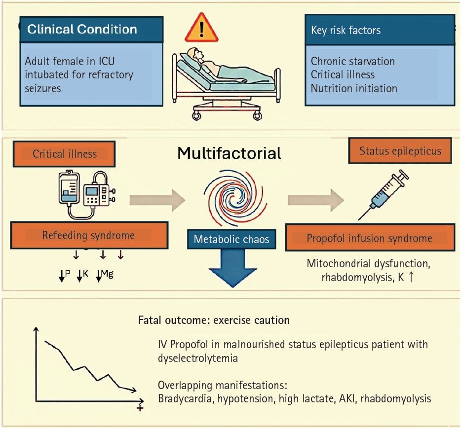
Metabolic factors contributing to clinical deterioration. ICU, intensive care unit; P, serum phosphorus; K, serum potassium; Mg, serum magnesium; IV, intravenous; AKI, acute kidney injury.
Fig. 2.
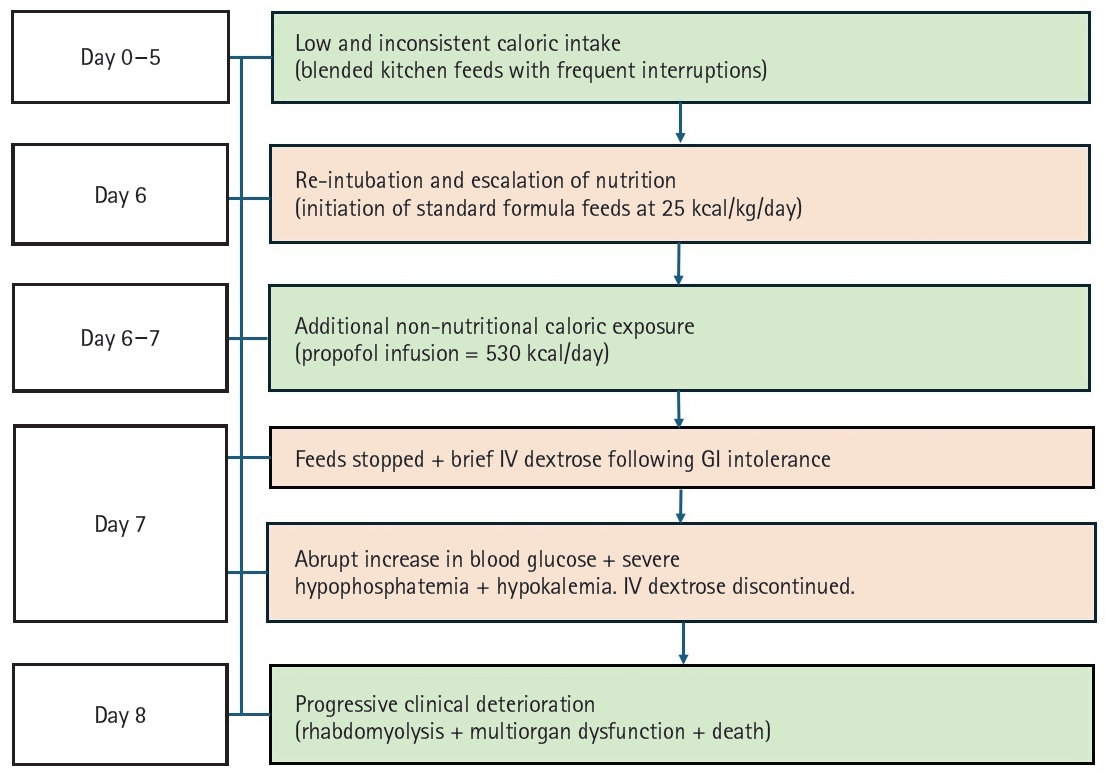
Timeline of nutritional exposure and metabolic deterioration. This schematic illustrates the temporal relationship between changes in nutritional intake, additional caloric exposure from propofol and dextrose, the development of electrolyte abnormalities, and subsequent clinical deterioration, supporting the diagnosis of refeeding syndrome in the setting of critical illness. IV, intravenous; GI, gastrointestinal.
Table 1.
Temporal progression of key metabolic parameters during ICU course
ICU, intensive care unit; K, serum potassium; P, serum phosphorus; Mg, serum magnesium; CPK, creatine phosphokinase; NR, not recorded; NG, nasogastric; AKI, acute kidney injury.
aDay 8 initial laboratory values were obtained at 4:00 AM. Propofol was stopped at 1:00 PM. Rhabdomyolysis, anuria, hyperkalemia, and cardiac arrest occurred at 10:00 PM.
- 1. da Silva JSV, Seres DS, Sabino K, Adams SC, Berdahl GJ, Citty SW, et al. ASPEN consensus recommendations for refeeding syndrome. Nutr Clin Pract 2020;35:178-95. ArticlePubMedPDF
- 2. Marik PE, Bedigian MK. Refeeding hypophosphatemia in critically ill patients in an intensive care unit: a prospective study. Arch Surg 1996;131:1043-7. ArticlePubMed
- 3. Dickerson RN, Buckley CT. Impact of propofol sedation upon caloric overfeeding and protein inadequacy in critically ill patients receiving nutrition support. Pharmacy (Basel) 2021;9:121.ArticlePubMedPMC
- 4. Charriere M, Ridley E, Hastings J, Bianchet O, Scheinkestel C, Berger MM, et al. Propofol sedation substantially increases the caloric and lipid intake in critically ill patients. Nutrition 2017;42:64-8. ArticlePubMed
- 5. Mirrakhimov AE, Voore P, Halytskyy O, Khan M, Ali AM. Propofol infusion syndrome in adults: a clinical update. Crit Care Res Pract 2015;2015:260385.ArticlePubMedPMCPDF
- 6. Hemphill S, McMenamin L, Bellamy MC, Hopkins PM. Propofol infusion syndrome: a structured literature review and analysis of published case reports. Br J Anaesth 2019;122:448-59. ArticlePubMedPMC
- 7. Hwang WS, Gwak HM, Seo DW. Propofol infusion syndrome in refractory status epilepticus. J Epilepsy Res 2013;3:21-7. ArticlePubMedPMC
- 8. Zhang Y, Qian M, Zheng A, Chen Y, Li B, Tang J, et al. The risk of propofol infusion syndrome on epilepsy patients: insights from FAERS data and published case reports. Eur J Pharmacol 2025;999:177429.ArticlePubMed
- 9. Walli A, Poulsen TD, Dam M, Borglum J. Propofol infusion syndrome in refractory status epilepticus: a case report and topical review. Case Rep Emerg Med 2016;2016:3265929.ArticlePubMedPMCPDF
- 10. Sobotka L. Basics in clinical nutrition: refeeding syndrome. E Spen Eur E J Clin Nutr Metab 2010;5:e146-7. Article
- 11. Crook MA. Refeeding syndrome: problems with definition and management. Nutrition 2014;30:1448-55. ArticlePubMed
- 12. Ha SW, Hong S. Recent advances in refeeding syndrome in critically ill patients: a narrative review. Ann Clin Nutr Metab 2024;16:3-9. Article
- 13. McKnight CL, Newberry C, Sarav M, Martindale R, Hurt R, Daley B, et al. Refeeding syndrome in the critically ill: a literature review and clinician's guide. Curr Gastroenterol Rep 2019;21:58.ArticlePubMedPDF
- 14. Ponzo V, Pellegrini M, Cioffi I, Scaglione L, Bo S. The refeeding syndrome: a neglected but potentially serious condition for inpatients. A narrative review. Intern Emerg Med 2021;16:49-60. ArticlePubMedPDF
References
Figure & Data
REFERENCES
Citations
Citations to this article as recorded by 
 Cite
Cite XML Download
Metabolic deterioration in a patient with refractory status epilepticus complicated by refeeding syndrome: a case report
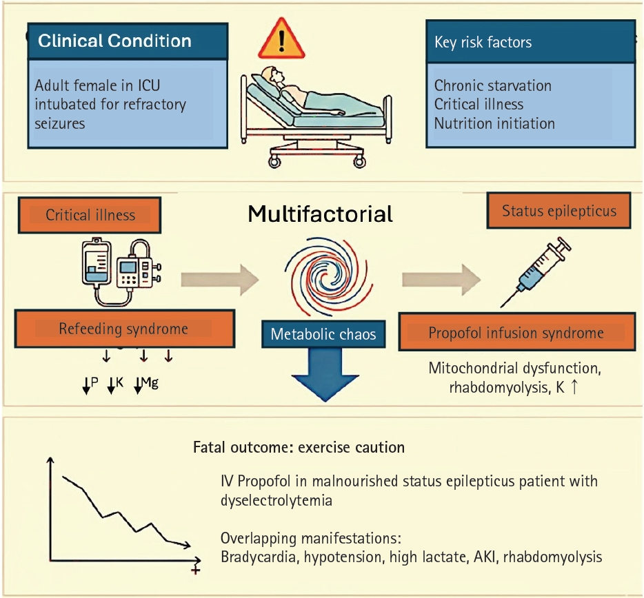
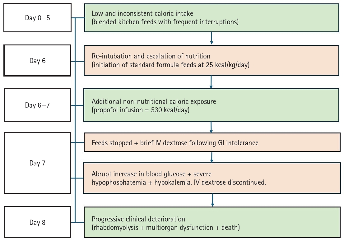
Fig. 1. Metabolic factors contributing to clinical deterioration. ICU, intensive care unit; P, serum phosphorus; K, serum potassium; Mg, serum magnesium; IV, intravenous; AKI, acute kidney injury.
Fig. 2. Timeline of nutritional exposure and metabolic deterioration. This schematic illustrates the temporal relationship between changes in nutritional intake, additional caloric exposure from propofol and dextrose, the development of electrolyte abnormalities, and subsequent clinical deterioration, supporting the diagnosis of refeeding syndrome in the setting of critical illness. IV, intravenous; GI, gastrointestinal.
Fig. 1.
Fig. 2.
Metabolic deterioration in a patient with refractory status epilepticus complicated by refeeding syndrome: a case report
| Hospital day | Key clinical events | `K (mEq/L) | P (mg/dL) | Mg (mg/dL) | Lactate (mmol/L) | CPK (IU/L) |
|---|---|---|---|---|---|---|
| Day 0 (Admission) | Seizure and intubation | 3.4 | 2.5 | 1.4 | 1.2 | NR |
| Day 1–3 | ICU stay, NG feeds (blended diet), extubation | 2.5–3.0 | NR | NR | NR | NR |
| Day 6 | Reintubation, commercial feeds started | 2.6 | 2.2 | 1.5 | 1.8 | NR |
| Day 7 | Propofol infusion (4 mg/kg/hr), hypotension, hypothermia, feed intolerance | 2.5 | 0.6 | 1.4 | 5.0 | 8,000 |
| Day 8a | Propofol stopped at 1:00 PM | 2.8 | 2.0 | 1.3 | 2.5 | - |
| Sudden collapse, AKI, cardiac arrest | 6.2 | 2.5 | NR | >14 | 80,000 |
Table 1. Temporal progression of key metabolic parameters during ICU course
ICU, intensive care unit; K, serum potassium; P, serum phosphorus; Mg, serum magnesium; CPK, creatine phosphokinase; NR, not recorded; NG, nasogastric; AKI, acute kidney injury. aDay 8 initial laboratory values were obtained at 4:00 AM. Propofol was stopped at 1:00 PM. Rhabdomyolysis, anuria, hyperkalemia, and cardiac arrest occurred at 10:00 PM.


