
 E-submission
 KSPEN
KSPEN KSSMN
KSSMN ASSMN
ASSMN JSSMN
JSSMNIndexed in:
Scopus, KCI, KoreaMed
Scopus, KCI, KoreaMed

Articles
- Page Path
- HOME > Ann Clin Nutr Metab > Volume 18(2); 2026 > Article
- Original Article Effects of oral deprivation and continuous gastrostomy feeding on masticatory function in rats
-
Airi Honjo
 , Ippei Yamaoka
 -
Annals of Clinical Nutrition and Metabolism 2026;18(2):173-180.
DOI: https://doi.org/10.15747/ACNM.26.0051
Published online: July 31, 2026
Medical Foods Research Institute, Otsuka Pharmaceutical Factory, Inc., Naruto, Japan
- Corresponding author: Ippei Yamaoka, email: yamaokai@otsuka.jp
• Received: March 13, 2026 • Revised: June 23, 2026 • Accepted: June 29, 2026
© 2026 The Korean Society of Surgical Metabolism and Nutrition · The Korean Society for Parenteral and Enteral Nutrition · Asian Society of Surgical Metabolism and Nutrition
This is an Open Access article distributed under the terms of the Creative Commons Attribution Non-Commercial License (http://creativecommons.org/licenses/by-nc/4.0), which permits unrestricted non-commercial use, distribution, and reproduction in any medium, provided the original work is properly cited.
- 575 Views
- 9 Download


Abstract
-
Purpose Tube feeding is frequently used in older adults and patients with dysphagia. However, when oral intake cannot be resumed, masticatory function may decline. Eating provides nutrition and helps maintain oral function, but the effects of different nutritional routes on masticatory muscles remain unclear. This study investigated the effects of a feeding model that combined differences in feeding route, diet form, and oral deprivation on masticatory-related muscles and masticatory function.
-
Methods Seven-week-old male Sprague–Dawley rats were assigned to receive a solid diet (solid group), a liquid diet (liquid group), or continuous gastric feeding via gastrostomy (gastrostomy group) for 3 weeks. The time required to complete feed intake, masseter electromyographic activity, and masticatory muscle weights were evaluated.
-
Results Energy intake and body weight changes did not differ significantly among the groups during the feeding period. Masseter muscle weight was significantly lower in the liquid group than in the other groups, whereas temporalis muscle weight tended to be higher in the solid group. The time required to complete feed intake was significantly longer in the gastrostomy group (389.7±210.4 sec/g) than in the solid and liquid groups (140.0±21.4 and 207.8±82.2 sec/g, respectively; P<0.01). When solid food was consumed after the 3-week feeding period, muscle activity during eating was higher in both the liquid and gastrostomy groups than in the solid group.
-
Conclusion Feeding models involving oral deprivation or gastrostomy feeding altered masticatory-related muscle characteristics and chewing performance in rats. Reduced feeding activity may contribute to impaired chewing and changes in tongue weight.
Graphical abstract
Introduction
Tube feeding is widely used for nutritional management in older adults and patients with reduced swallowing function. Among tube feeding methods, nasogastric tube feeding and percutaneous endoscopic gastrostomy are commonly used to administer enteral nutrition [1]. Tube feeding is therefore a useful option for nutritional management. However, some individuals have difficulty transitioning from tube feeding to oral intake, and declines in masticatory and feeding function are major concerns [2].
Diet provides energy and nutrients and also helps maintain oral functions, including mastication and swallowing. Even when diets have the same energy content, intake through the oral cavity may have distinct physiological and psychological significance [3]. Masticatory movement is achieved through coordinated activity of the masseter and temporalis muscles and the tongue, and activity generated during food consumption is essential for maintaining masticatory function. During mastication, the masseter muscle primarily controls jaw-closing force, whereas the temporalis muscle helps control occlusal force and mandibular movement [4]. The tongue also plays an essential role in feeding, including bolus formation and food transport [5]. Because these masticatory-related muscles contribute directly to oral function, their weight is closely related to masticatory function, and decreased frequency and intensity of masticatory activity can lead to muscle atrophy and functional decline [6]. These findings suggest that reduced oral motor activity and oral stimulation during prolonged tube feeding may be associated with alterations in masticatory muscle function.
Previous animal studies have evaluated the effects of different food forms on masticatory function and intake behavior [7-10]. For example, rodent studies have compared solid feed with powder or liquid feed, which induces less mastication [11-13]. These studies have demonstrated the role of adequate mastication in maintaining masticatory muscle function, even when nutrition is sufficient. However, tube feeding–induced declines in masticatory muscle function have not been adequately reproduced in experimental animals, and no studies have examined the role of oral nutritional intake, compared with tube feeding, in preserving masticatory function.
In the present study, some rats received continuous intragastric infusion of liquid feed that bypassed oral intake, whereas others received solid or liquid feed orally with the same nutritional composition. This design allowed comparison of feeding models involving oral solid feeding, oral liquid feeding, and continuous gastrostomy feeding. We hypothesized that the absence of oral intake would lead to functional decline in rat masticatory muscles. To test this hypothesis, we examined the significance of oral intake in tube-fed rats using objective indicators, including the time required to complete solid-feed intake and the weight of the masticatory muscles.
Methods
All procedures were approved by Otsuka Pharmaceutical Factory, Inc. (approval number: OPFCAE-2023184), and were conducted in accordance with the animal experimentation guidelines of Otsuka Pharmaceutical Factory, Inc., which comply with relevant Japanese regulations and guidelines for the care and use of laboratory animals, including the Act on Welfare and Management of Animals and the Fundamental Guidelines for Proper Conduct of Animal Experiments and Related Activities. This study is reported in accordance with the ARRIVE guidelines.
A harm–benefit analysis was performed to justify the experimental design, including 3R measures: reduced sample size, pilot studies, intervention criteria, and environmental enrichment. The endpoint fate of all animals was documented. Animals were monitored daily throughout the study for general health status, body weight, food intake, catheter condition, wound healing, and signs of pain or distress. Humane endpoints were established before study initiation. Animals showing severe deterioration in health status or complications associated with the gastrostomy catheter were removed from the study and euthanized according to the predefined humane endpoint criteria. The sample size was determined on the basis of previous experience with similar exploratory animal studies and practical considerations rather than a formal statistical power calculation.
The purchased animals were allowed to acclimate, during which AIN-93G solid feed (Oriental Yeast Co., Ltd.) was provided ad libitum. After the acclimation period, AIN-93G feed was replaced with the test feed. To prepare the test feed, a liquid food (HINEX E-Gel, Otsuka Pharmaceutical Factory, Inc.) was freeze-dried into powder and then solidified using a feed manufacturing machine (Oriental Yeast Co., Ltd.). The composition of the test feed is shown in Table 1.
Thirty 7-week-old male Crl:CD Sprague–Dawley rats (Jackson Laboratory Japan, Inc., Kanagawa, Japan) were used. The rats were housed at 23±3 °C and 55%±15% humidity under a 12-hour light/dark cycle, with lights on from 7:00 to 19:00.
Feeding with the test feed was initiated after the 5-day acclimation period. Animals were then allocated to three experimental groups using body weight–based allocation to ensure comparable baseline body weights among groups: the solid group, which received oral intake of feed prepared by solidifying a liquid food; the liquid group, which received oral intake of a liquid food; and the gastrostomy group, which received liquid food through a gastrostomy catheter. Rats in the gastrostomy group were fasted from the day before group allocation. Laparotomy was subsequently performed under isoflurane anesthesia, and a catheter was placed in the stomach, as described previously [14]. After surgery, the rats received intramuscular ampicillin preparation and were monitored daily for general condition, body weight, food intake, wound healing, and catheter patency. Humane endpoints had been predefined before study initiation, and animals with severe complications associated with the gastrostomy catheter or other signs of substantial distress were removed from the study and euthanized according to institutional guidelines.
The rats were housed individually from the day of group allocation, and the test-feed intake period was set at 3 weeks. The solid group was offered test feed providing 80 kcal/day, and actual intake was determined by subtracting the amount of unconsumed feed collected daily. The liquid and gastrostomy groups were pair-fed on the basis of the actual intake of the solid group measured on the previous day. For the liquid group, the test feed was poured into a glass feeding dish, placed inside the cage, and changed daily. For the gastrostomy group, the test feed was loaded into a syringe (J-Feed injector, JMS Co., Ltd.), connected to the upper end of the animal-specific gastrostomy catheter, and infused continuously over 24 hours using a syringe pump. In the liquid and gastrostomy groups, access to drinking water was discontinued after test-feed intake began, and the rats received only water contained in the test feed.
After 3 weeks of test-feed intake, the rats underwent surgery to place a transmitter for measuring muscle potential. The transmitter, receiving board, and data acquisition system were components of the long-term automatic telemetry measurement system (Data Sciences International [DSI]). Under isoflurane anesthesia, laparotomy was performed in five rats per group, and the transmitter was placed in the peritoneal cavity. Because telemetry devices were limited, five animals from each group were selected for electromyographic measurements. Animals were selected on the basis of body weight to ensure representative body-weight distributions within each group. After the abdominal procedure, an incision was made on the right side of the face, and a recording needle electrode was inserted into the superficial layer of the right masseter muscle and fixed with sutures. After the facial surgical procedure, the rats received intramuscular ampicillin preparation and were awakened from anesthesia. During recovery, the animals were monitored daily for postoperative health status, wound healing, body condition, and signs of pain or distress. After the surgical procedures, a 2-day recovery period was established, during which the rats were fasted.
After the recovery period, each group was administered 2.5 g of the specified test feed, and muscle activity generated until feed intake was completed was measured. The time required to complete feed intake was also measured. If no feeding behavior was observed within the maximum measurement time of 1.5 hours, measurement was discontinued, and the remaining feed was weighed to calculate the time required to consume 1 g of feed. After muscle activity was measured, all rats underwent tissue collection as follows. Laparotomy was performed under deep isoflurane anesthesia, and the rats were euthanized by exsanguination via incision of the abdominal vena cava. After death was confirmed, the masseter and temporalis muscles and tongue were removed from the left side of the face. The gastrocnemius and anterior tibial muscles were removed from the left lower limb and weighed using a precision electronic balance. The masseter muscle was treated as a single sample without separating the superficial and deep layers.
Outcome assessments were conducted without blinding to group allocation.
Masseter muscle potential during feeding was analyzed using PONEMAH software (DSI), which was designed for a long-term automatic telemetry measurement system. Muscle-potential data were aggregated only during periods of mastication activity, and data were excluded using a peak threshold of 0.05 mV·s. The sum of muscle potentials during mastication was calculated as the amount of muscle activity.
All results are expressed as mean±standard deviation. Before analysis of variance (ANOVA), normality was assessed using the Shapiro–Wilk test, and homogeneity of variance was assessed using Levene’s test. All outcome measures satisfied the assumptions of normality and homogeneity of variance (all P>0.05). Statistical analyses were conducted using one-way ANOVA. When a significant overall group effect was observed, Fisher’s least significant difference test was used for post hoc pairwise comparisons. Fisher’s least significant difference test was selected because this study was exploratory and involved only three predefined comparison groups. The significance level was set at 5%. All analyses were performed using Excel Statistics, version 3.20 (Social Survey Research Information Co., Ltd.).
Results
The flow of animals through the study, including exclusions and analysis populations, is summarized (Fig. 1). The three groups did not differ significantly in total energy intake or body weight at autopsy (Table 2). Throughout the test-feed intake period, the general condition of rats in the solid and liquid groups was favorable. Three rats in the gastrostomy group met the predefined humane endpoint criteria because of gastrostomy catheter obstruction or catheter dislodgement from the stomach; these rats were euthanized and excluded from further analyses. In addition, one rat each in the solid and liquid groups was excluded from the feeding-time analysis because feeding behavior could not be reliably recorded. No substantial violations of normality or homogeneity of variance were detected.
Masseter muscle weight was significantly lower in the liquid group than in the solid and gastrostomy groups (Fig. 2A). Temporalis muscle weight tended to be lower in the liquid and gastrostomy groups than in the solid group, although the overall group effect did not reach statistical significance (ANOVA: F(2,24)=3.09, P=0.064) (Fig. 2B). Consistent with this finding, Tukey-adjusted post hoc analyses did not identify statistically significant differences among groups. Tongue weight was significantly lower in the gastrostomy group than in the other groups (Fig. 2C). In contrast, gastrocnemius and anterior tibial muscle weights did not differ significantly among the three groups (Fig. 2D and E).
The time required to complete intake of masticatory solid feed was significantly longer in the gastrostomy group than in the other groups (Fig. 3). Masseter muscle potential during intake of masticatory solid feed was significantly higher in the liquid and gastrostomy groups than in the solid group (Fig. 4).
Discussion
Mastication, the first major step in digestion, requires coordination of the tongue, facial muscles, jaw, and teeth. Long-term administration of enteral nutrition may be associated with poor systemic health and gradual development of masticatory and swallowing dysfunction, indicating that maintaining the masticatory system is important for tube-fed patients [2]. Therefore, experimental animals that simulate the clinical conditions of tube-fed patients should be investigated. Previous animal studies have compared the effects of different feed forms [11-13,15], but no studies have comparatively evaluated the effects of tube feeding. In the present study, rats received nutrition through continuous intragastric infusion of liquid feed without mastication and were compared with rats receiving oral intake of solid or liquid feed with the same nutritional composition.
Masticatory muscle activity is influenced by the physical properties of food. In rabbits, soft feed has been shown to decrease the number of daily bursts and the activity time of the masseter muscle [12], and similar results have been reported in rats fed powder feed [11]. On the basis of these findings, we hypothesized that restricted masticatory movement may impair masticatory function. To test this hypothesis, we compared the amount of muscle activity (mV·s) in rats, as determined from the waveform of the masseter muscle potential. The amount of muscle activity generated until completion of solid-feed intake was significantly higher in the liquid and gastrostomy groups than in the solid group. Increased masticatory muscle activity during consumption of food with the same physical properties may reflect reduced mastication efficiency; therefore, the higher total muscle activity observed in the liquid and gastrostomy groups was likely attributable to restricted masticatory movement in the present study. The time required to complete feed intake was also longest in the gastrostomy group, suggesting that feeding behavior was affected by disuse of oral function. Because the gastrostomy group received no oral intake during nutrition administration, the longer feeding time may reflect changes in the coordinated use of oral structures involved in mastication and swallowing, requiring compensatory muscle activity when solid-feed intake resumed. In addition, tongue weight was significantly decreased only in the gastrostomy group, which may reflect reduced tongue muscle mass associated with prolonged absence of oral feeding activity.
This reduction may have contributed to decreased ability to form and transport a bolus. A study examining the relationship between tongue pressure and masticatory function reported that reduced tongue pressure decreased the ability to form a bolus and delayed food clearance from the oral cavity [16]. These findings suggest that decreased tongue weight may hinder bolus formation and swallowing movement and, in the present study, may have contributed to the prolonged time required to complete solid-feed intake in the gastrostomy group. In the liquid group, continuous oral stimulation likely contributed to partial maintenance of basic oral-movement functions, such as the swallowing reflex and bolus transport, although no mastication load was present during the feeding period. The liquid group did not differ significantly from the solid group in the time required to complete solid-feed intake, despite exhibiting greater muscle activity. This finding suggests that increased muscle activity in the liquid group may have compensated for slightly reduced masticatory efficiency during solid-feed consumption, thereby maintaining the time required to complete feed intake. Overall, these observations suggest an association between reduced oral feeding activity and changes in chewing performance and masticatory-related muscles.
Inefficient feeding movement may therefore increase muscle activity and slow the rate of food intake.
Next, we focused on the weight of masticatory muscles in rats receiving no oral feed intake, hypothesizing that the most prominent weight changes would occur in muscles with major roles in mastication. To test this hypothesis, we measured the weights of tissues involved in mastication: the masseter muscle, temporalis muscle, and tongue. Masseter muscle weight was significantly lower in the liquid group than in the other groups, with no significant difference between the gastrostomy and solid groups. Temporalis muscle weight tended to be lower in the liquid and gastrostomy groups. Previous studies have reported functional alterations in masticatory muscles in animals fed diets requiring reduced mastication [11,13]. Because masticatory efficiency is improved by coordinated activity of the masseter and temporalis muscles, decreased weight of either muscle may contribute to reduced masticatory efficiency [4]. Decreased masticatory muscle weight may also affect masticatory rhythm and muscle coordination. The temporalis muscle, which works with the masseter muscle to increase masticatory efficiency, has an important role in the power phase [4]. Although temporalis muscle weight tended to be lower in the liquid and gastrostomy groups, the overall group effect did not reach statistical significance (ANOVA: F(2,24)=3.09, P=0.064). Therefore, any contribution of temporalis muscle changes to the observed differences in chewing performance remains speculative. However, masseter muscle weight in the gastrostomy group was comparable to that in the solid group (Fig. 2A). Botulinum toxin A (BoNTA) injection into the unilateral masseter muscle induced molecular and morphological changes related to muscle atrophy and affected the entire ipsilateral masticatory musculature [17]. These reports suggest that functional impairment of some masticatory muscles may broadly affect other masticatory muscles.
A possible explanation for the absence of decreased masseter muscle weight in the gastrostomy group is compensatory change associated with muscle degeneration. Previous studies using a model of long-term disuse induced by BoNTA have reported increases in connective tissue, collagen content, and fatty infiltration in the masseter muscle [18]. However, because no histological analyses were performed in the present study, the involvement of such tissue changes remains speculative and should be interpreted with caution. Further studies including histological evaluation are needed to clarify this mechanism. In contrast, in the liquid group, muscle degeneration may have progressed more gradually because the rats maintained some level of oral intake, albeit limited, which may have reduced muscle weight without compensatory tissue changes. Notably, despite preserved muscle weight in the gastrostomy group, functional impairment was suggested by increased muscle activity and prolonged feeding time. Disuse of the gastrocnemius muscle, which shares fast-twitch characteristics with the masseter muscle, has been reported to increase the proportion of connective tissue [19], supporting this proposed mechanism. Further studies are warranted to better characterize the underlying mechanism.
The results of this study suggest that prolonged non-oral feeding may be associated with reduced function of the masticatory muscles and tongue. Our findings also suggest that daily masticatory movement and oral stimulation are important for maintaining function. In contrast, continued oral intake in the liquid group, although the feed was liquid, may have suppressed declines in masticatory muscle weight and function to some extent. These findings provide preliminary insight into potential changes in oral function associated with prolonged non-oral feeding. However, direct extrapolation to older adults, patients with dysphagia, or long-term enterally fed patients should be made cautiously.
The present study had several limitations. First, rats in the gastrostomy group differed from those in the solid and liquid groups, which were exposed to feed for a certain period. Therefore, the possibility that differences in acclimation to feed, in addition to decreased masticatory muscle function, contributed to the prolonged time required to complete feed intake cannot be completely ruled out. Second, the time required to complete feed intake was assessed at only a single time point, and the subsequent recovery process was not investigated. Third, the 48-hour fasting period after transmitter implantation may have affected muscle volume and function in these rats through metabolic changes and weight loss. This fasting period was used to allow adequate recovery by avoiding potential effects of feeding on the wound site during the early postoperative period. However, the possibility that fasting affected the results of the feeding intervention cannot be completely ruled out. Fourth, although the gastrostomy group underwent laparotomy and catheter placement, sham surgery was not performed in the other groups. Therefore, the possibility that surgical invasiveness affected some of the observed changes cannot be excluded. Fifth, this study focused on masticatory function and did not include functional evaluation of swallowing coordination. Swallowing-related muscle activity was not measured because of technical complexity and low reproducibility. The absence of evaluations of swallowing reflex and bolus transport efficiency is a key limitation of this study. In light of these limitations, future research should include comprehensive evaluation with histological analysis and swallowing-function assessment.
The present findings suggest that prolonged reduction or absence of oral feeding activity may affect masticatory-related muscles and chewing performance. Continuous oral stimulation, even with a liquid diet, appeared to partially preserve oral function compared with complete bypass of oral feeding through gastrostomy. Further studies are needed to clarify the underlying mechanisms and their relevance to clinical populations requiring long-term enteral nutrition.
Authors’ contribution
Conceptualization: IY. Methodology: AH, IY. Formal analysis: AH, IY. Validation: AH, IY. Project administration: IY. Writing–original draft: AH. Writing–review and editing: AH, IY. All authors read and approved of the final manuscript.
Conflict of interest
Airi Honjo and Ippei Yamaoka are employees of Otsuka Pharmaceutical Factory, Inc. Otherwise, no other potential conflict of interest relevant to this article was reported.
Funding
None.
Data availability
Contact the corresponding author for research data availability.
Acknowledgments
The authors would like to thank Dr. Tomohiro Kagawa, and Ms. Erika Mori (Otsuka Pharmaceutical Factory, Inc.), who provided dedicated technical support for the animal experiments.
Supplementary materials
None.
Fig. 1.
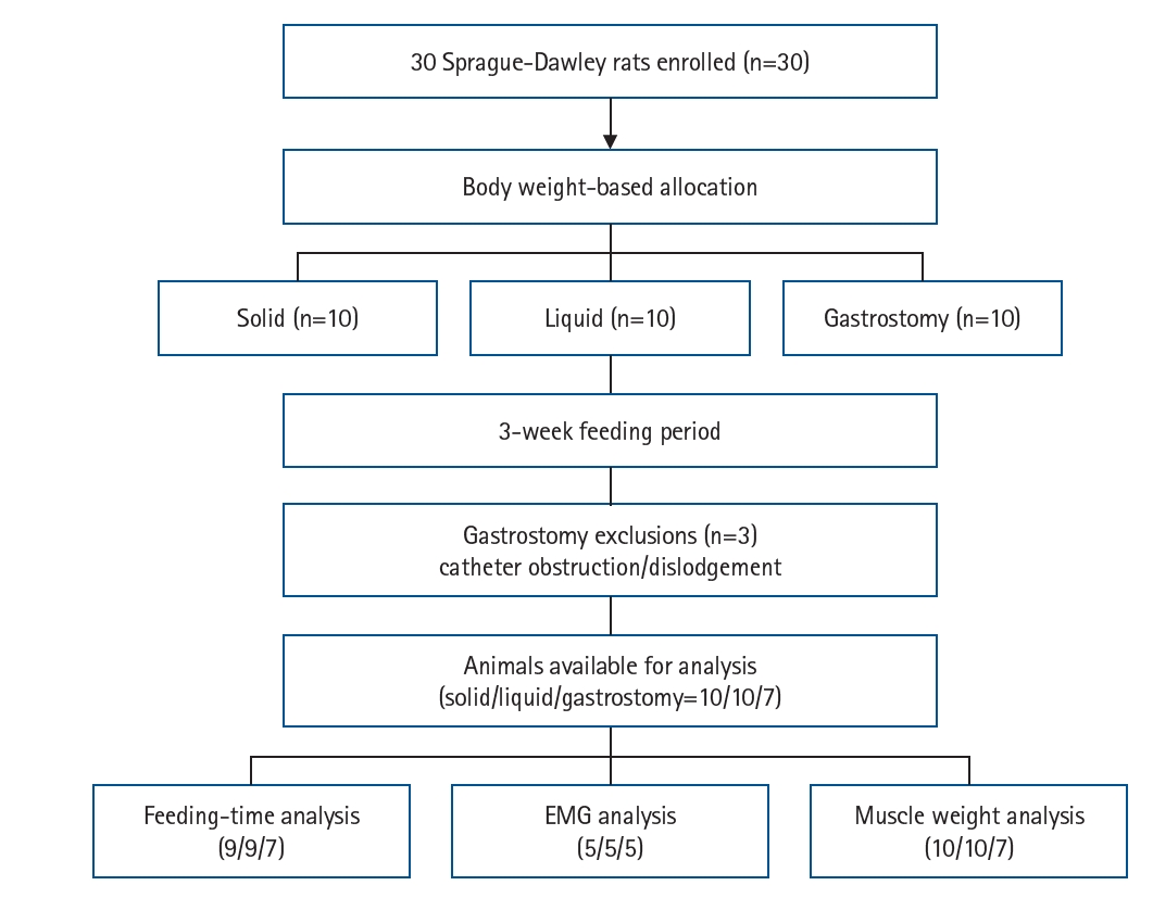
Animal flow diagram prepared according to the ARRIVE 2.0 guidelines. Thirty rats were allocated to experimental groups using body weight–based allocation. Three rats in the gastrostomy group met predefined humane endpoint criteria because of catheter obstruction or dislodgement and were excluded from further analyses. One rat each in the solid and liquid groups was excluded from the feeding-time analysis because feeding behavior could not be reliably recorded. Five animals per group were selected for electromyographic analysis because of the limited availability of telemetry devices. EMG, electromyography.
Fig. 2.
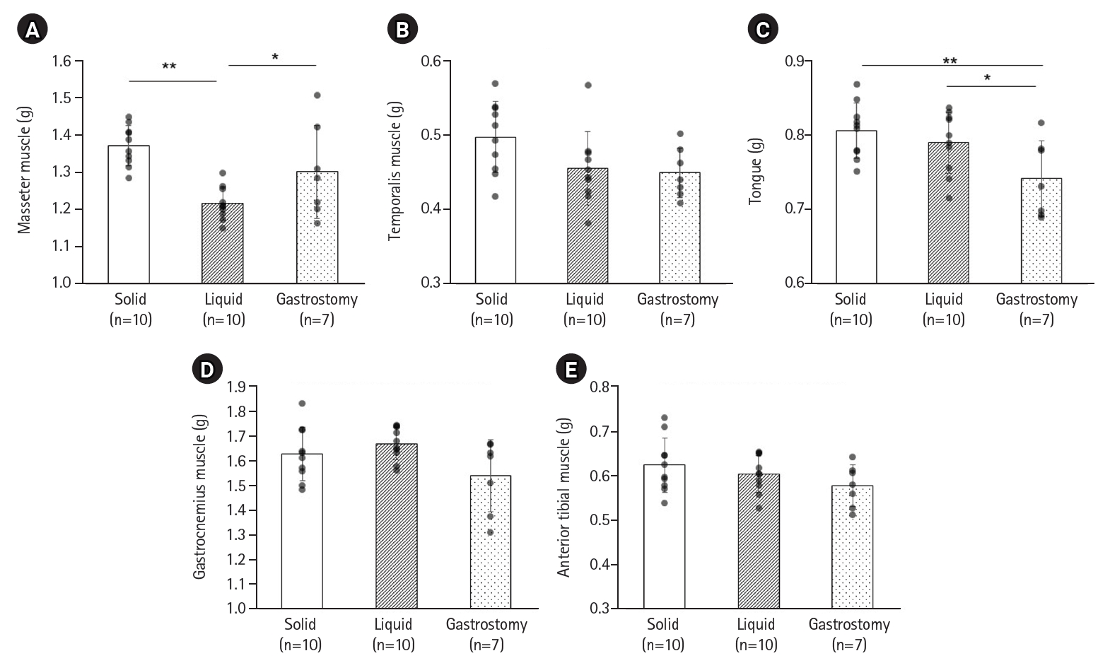
Weights of masticatory muscles and lower-limb skeletal muscles. (A) Masseter muscle, (B) temporalis muscle, (C) tongue, (D) gastrocnemius muscle, and (E) anterior tibial muscle. Data are presented as mean±standard deviation. *P<0.05, **P<0.01 (Fisher’s least significant difference test).
Fig. 3.
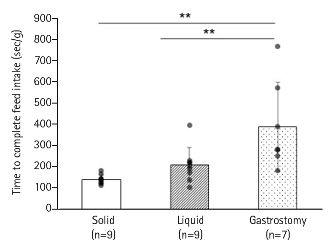
Time required to complete intake of the provided solid feed. One animal each in the solid and liquid groups was excluded from this analysis because feeding behavior could not be reliably recorded. Data are presented as mean±standard deviation. **P<0.01 (Fisher's least significant difference test).
Fig. 4.
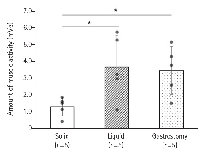
Masseter muscle activity during intake of solid feed. Data are presented as mean±standard deviation. *P<0.05 (Fisher's least significant difference test).
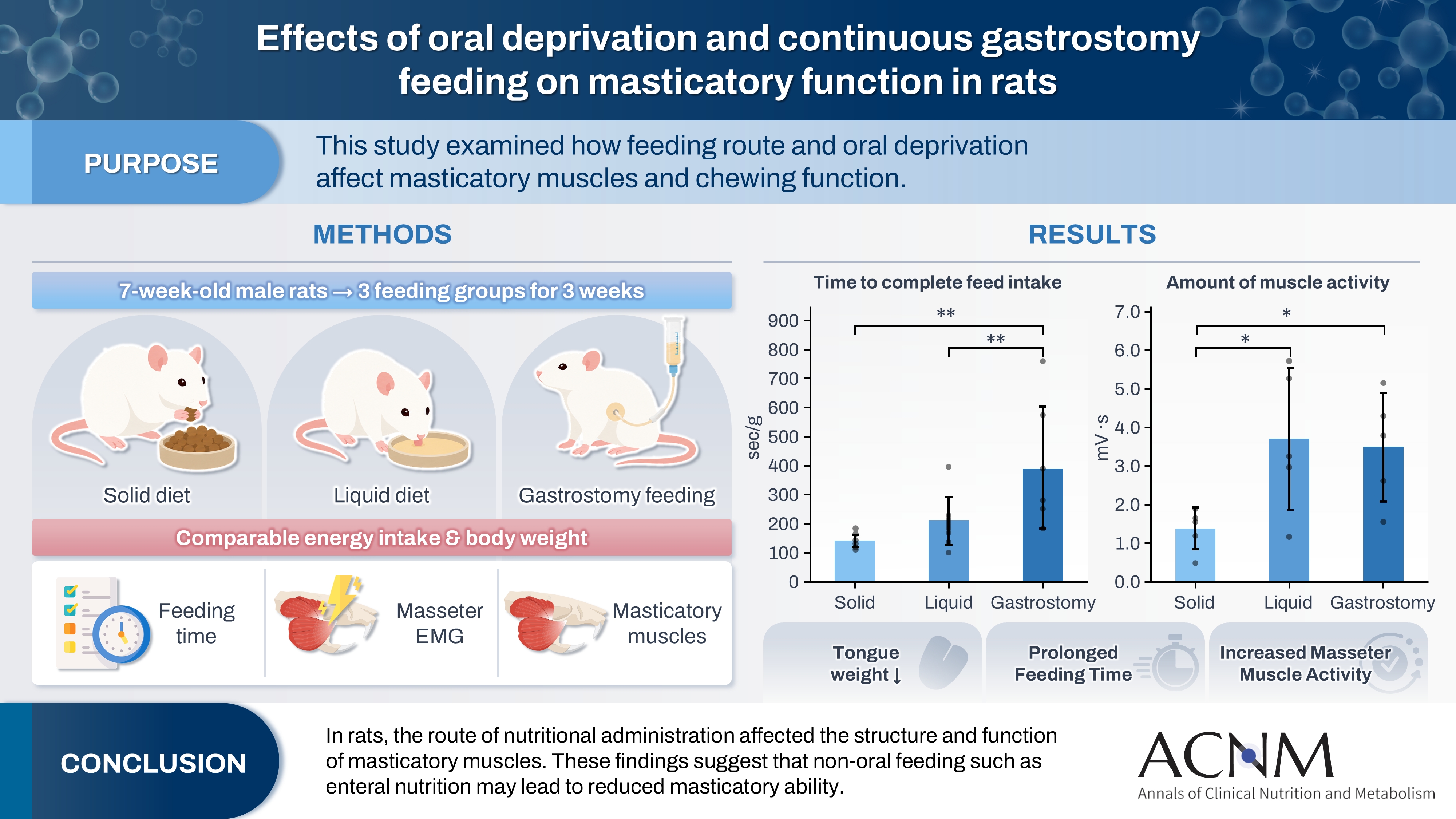
Table 1.
Nutritional composition of the test feed
| Group | Solid (n=10) | Liquid (n=10) | Gastrostomy (n=7) |
|---|---|---|---|
| Form | Solid | Liquid | Liquid |
| Energy (kcal/100 g) | 396a | 80 | 80 |
| Water (g/100 g) | 5.8 | 81.1 | 81.1 |
| Protein (g/100 g) | 15.8 | 4.0 | 4.0 |
| Fat (g/100 g) | 8.7 | 2.2 | 2.2 |
Table 2.
Body weight at autopsy and total energy intake
| Group | Solid (n=10) | Liquid (n=10) | Gastrostomy (n=7) | P-value |
|---|---|---|---|---|
| Final body weight (g) | 305.4±17.8 | 305.1±6.3 | 292.6±10.4 | 0.097 |
| Total energy intake (kcal) | 1,305.1±195.4 | 1,256.7±62.7 | 1,255.3±7.9 | 0.626 |
- 1. Ogita M, Utsunomiya H, Akishita M, Arai H. Indications and practice for tube feeding in Japanese geriatricians: implications of multidisciplinary team approach. Geriatr Gerontol Int 2012;12:643-51. ArticlePubMed
- 2. Furuya H, Kikutani T, Igarashi K, Sagawa K, Yajima Y, Machida R, et al. Effect of dysphagia rehabilitation in patients receiving enteral nutrition at home nursing care: a retrospective cohort study. J Oral Rehabil 2020;47:977-82. PubMedPMC
- 3. Terech-Skora S, Kasprzyk-Mazur J, Leyk-Kolanczak M, Kruk A, Piotrkowska R, Medrzycka-Dabrowska W, et al. Assessment of oral health in long-term enteral and parenteral nutrition patients: Significant aspects of nursing care. Int J Environ Res Public Health 2023;20:3381.ArticlePubMedPMC
- 4. Yoshimi T, Koga Y, Nakamura A, Fujishita A, Kohara H, Moriuchi E, et al. Mechanism of motor coordination of masseter and temporalis muscles for increased masticatory efficiency in mice. J Oral Rehabil 2017;44:363-74. ArticlePubMedPDF
- 5. Matsuo K, Palmer JB. Anatomy and physiology of feeding and swallowing: normal and abnormal. Phys Med Rehabil Clin N Am 2008;19:691-707. ArticlePubMedPMC
- 6. Liu ZZ, Rafferty KL, Wang DB, Owart B, Herring SW. Bilateral treatment of the masseter with botulinum toxin: consequences for mastication, muscle force and the mandibular condyle. J Oral Rehabil 2023;50:775-81. ArticlePubMed
- 7. Kakizaki Y, Uchida K, Yamamura K, Yamada Y. Coordination between the masticatory and tongue muscles as seen with different foods in consistency and in reflex activities during natural chewing. Brain Res 2002;929:210-7. ArticlePubMed
- 8. Tanaka E, Sano R, Kawai N, Langenbach GE, Brugman P, Tanne K, et al. Effect of food consistency on the degree of mineralization in the rat mandible. Ann Biomed Eng 2007;35:1617-21. ArticlePubMedPDF
- 9. Inoue M, Harasawa Y, Yamamura K, Ariyasinghe S, Yamada Y. Effects of food consistency on the pattern of extrinsic tongue muscle activities during mastication in freely moving rabbits. Neurosci Lett 2004;368:192-6. ArticlePubMed
- 10. Langenbach G, van de Pavert S, Savalle W, Korfage H, van Eijden T. Influence of food consistency on the rabbit masseter muscle fibres. Eur J Oral Sci 2003;111:81-4. ArticlePubMedPDF
- 11. Kawai N, Sano R, Korfage JA, Nakamura S, Kinouchi N, Kawakami E, et al. Adaptation of rat jaw muscle fibers in postnatal development with a different food consistency: an immunohistochemical and electromyographic study. J Anat 2010;216:717-23. ArticlePubMedPMC
- 12. Grunheid T, Brugman P, Zentner A, Langenbach GE. Changes in rabbit jaw-muscle activity parameters in response to reduced masticatory load. J Exp Biol 2010;213:775-81. ArticlePubMedPDF
- 13. Liu ZJ, Ikeda K, Harada S, Kasahara Y, Ito G. Functional properties of jaw and tongue muscles in rats fed a liquid diet after being weaned. J Dent Res 1998;77:366-76. ArticlePubMedPDF
- 14. Kagawa T, Yamaoka I. Intragastric infusion of a liquid diet with low-methoxyl pectin alleviates fecal inconsistency and local proinflammatory cytokine expression in lipopolysaccharide-septic rats. Nutrition 2024;118:112271.ArticlePubMed
- 15. Kono K, Tanikawa C, Murata Y, Yanagita T, Kamioka H, Yamashiro T, et al. Three-dimensional changes in the craniofacial complex associated with soft-diet feeding. Eur J Orthod 2020;42:509-16. ArticlePubMedPDF
- 16. Lee JH, Kim HS, Yun DH, Chon J, Han YJ, Yoo SD, et al. The relationship between tongue pressure and oral dysphagia in stroke patients. Ann Rehabil Med 2016;40:620-8. ArticlePubMedPMCPDF
- 17. Balanta-Melo J, Eyquem-Reyes A, Blanco N, Vasquez W, Kupczik K, Toro-Ibacache V, et al. Unilateral hypofunction of the masseter leads to molecular and 3D morphometric signs of atrophy in ipsilateral agonist masticatory muscles in adult mice. Int J Mol Sci 2023;24:14740.ArticlePubMedPMC
- 18. Ramos DM, de Brito Silva R, De la Torre Canales G, Resende L, Esquisatto MAM, Moreira NCF, et al. Histomorphometric changes of the masseter muscle of rats after a single injection of botulinum toxin type A. Aesthetic Plast Surg 2024;48:1037-44. ArticlePubMedPDF
- 19. Jarvinen TA, Jozsa L, Kannus P, Jarvinen TL, Jarvinen M. Organization and distribution of intramuscular connective tissue in normal and immobilized skeletal muscles: an immunohistochemical, polarization and scanning electron microscopic study. J Muscle Res Cell Motil 2002;23:245-54. ArticlePubMedPDF
References
Figure & Data
REFERENCES
Citations
Citations to this article as recorded by 
 Cite
Cite XML Download
Effects of oral deprivation and continuous gastrostomy feeding on masticatory function in rats
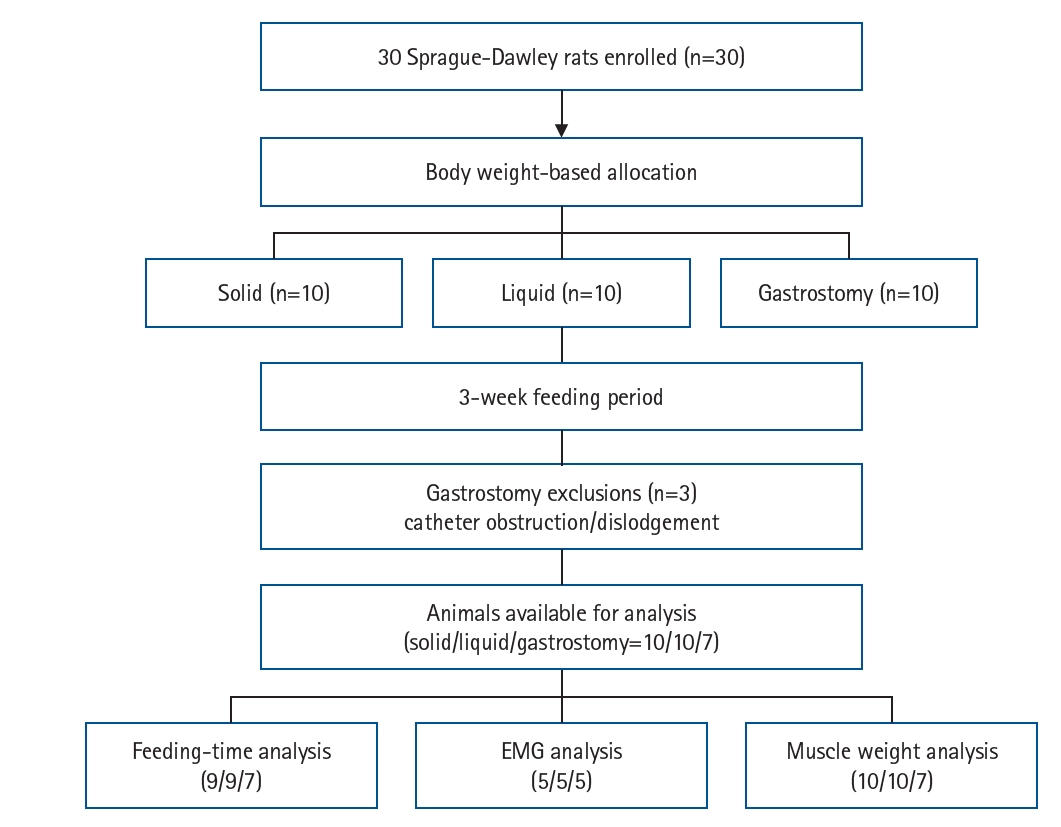
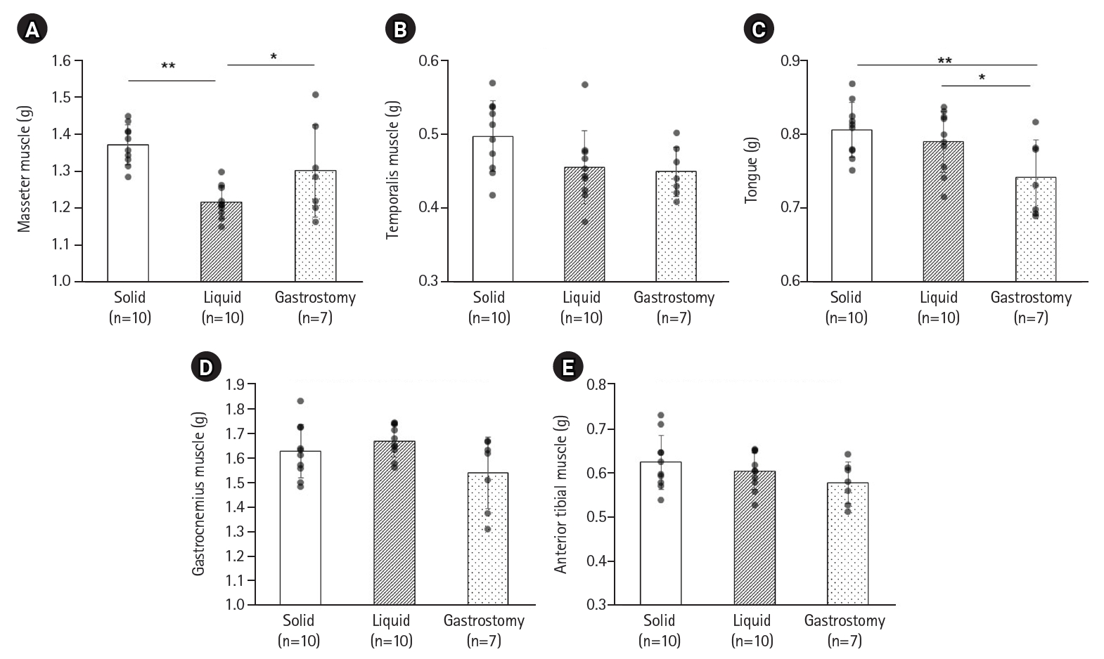
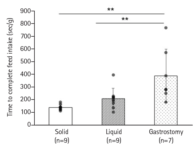
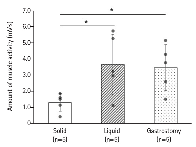
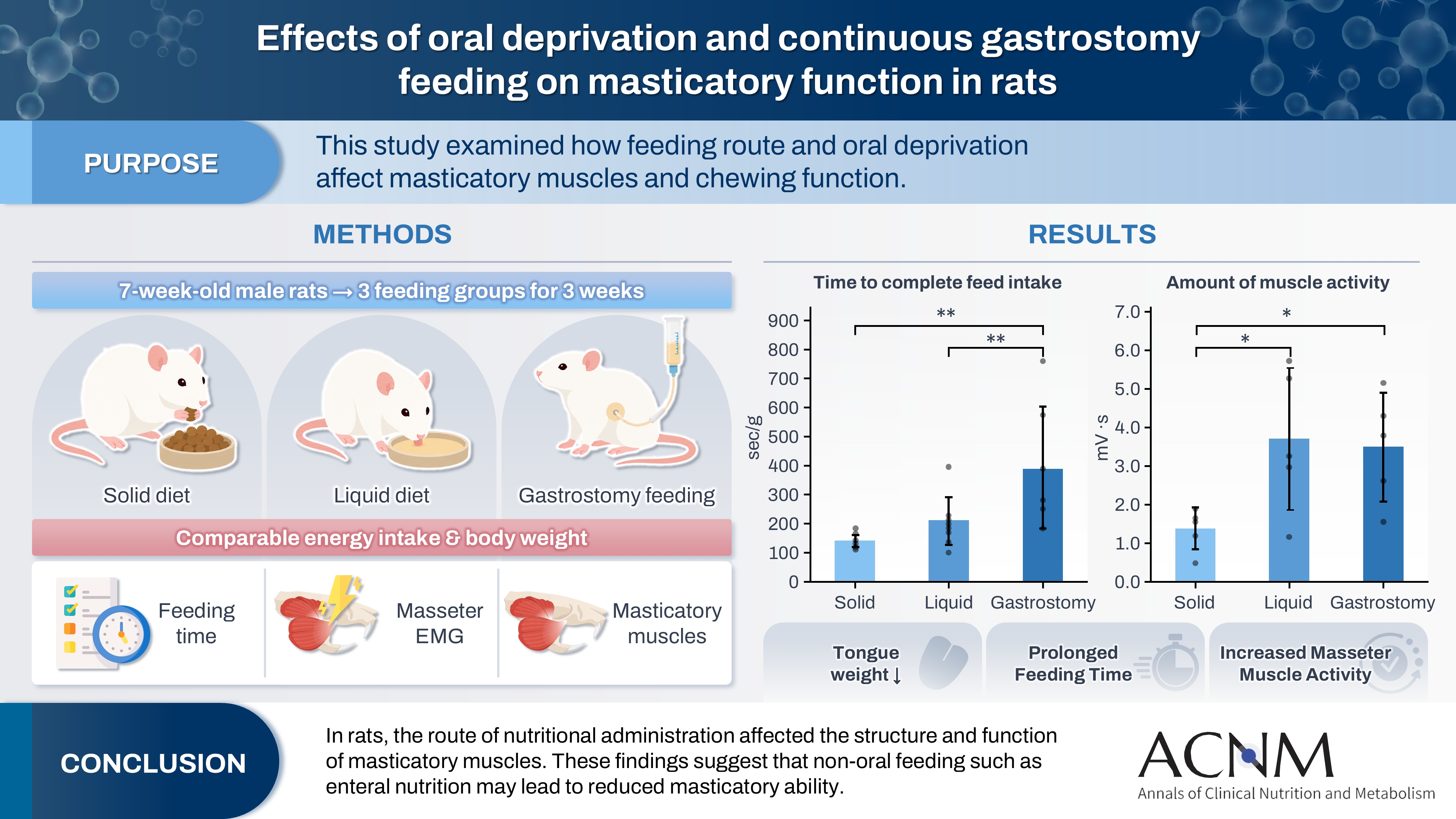
Fig. 1. Animal flow diagram prepared according to the ARRIVE 2.0 guidelines. Thirty rats were allocated to experimental groups using body weight–based allocation. Three rats in the gastrostomy group met predefined humane endpoint criteria because of catheter obstruction or dislodgement and were excluded from further analyses. One rat each in the solid and liquid groups was excluded from the feeding-time analysis because feeding behavior could not be reliably recorded. Five animals per group were selected for electromyographic analysis because of the limited availability of telemetry devices. EMG, electromyography.
Fig. 2. Weights of masticatory muscles and lower-limb skeletal muscles. (A) Masseter muscle, (B) temporalis muscle, (C) tongue, (D) gastrocnemius muscle, and (E) anterior tibial muscle. Data are presented as mean±standard deviation. *P<0.05, **P<0.01 (Fisher’s least significant difference test).
Fig. 3. Time required to complete intake of the provided solid feed. One animal each in the solid and liquid groups was excluded from this analysis because feeding behavior could not be reliably recorded. Data are presented as mean±standard deviation. **P<0.01 (Fisher's least significant difference test).
Fig. 4. Masseter muscle activity during intake of solid feed. Data are presented as mean±standard deviation. *P<0.05 (Fisher's least significant difference test).
Graphical abstract
Fig. 1.
Fig. 2.
Fig. 3.
Fig. 4.
Graphical abstract
Effects of oral deprivation and continuous gastrostomy feeding on masticatory function in rats
| Group | Solid (n=10) | Liquid (n=10) | Gastrostomy (n=7) |
|---|---|---|---|
| Form | Solid | Liquid | Liquid |
| Energy (kcal/100 g) | 396a | 80 | 80 |
| Water (g/100 g) | 5.8 | 81.1 | 81.1 |
| Protein (g/100 g) | 15.8 | 4.0 | 4.0 |
| Fat (g/100 g) | 8.7 | 2.2 | 2.2 |
| Group | Solid (n=10) | Liquid (n=10) | Gastrostomy (n=7) | P-value |
|---|---|---|---|---|
| Final body weight (g) | 305.4±17.8 | 305.1±6.3 | 292.6±10.4 | 0.097 |
| Total energy intake (kcal) | 1,305.1±195.4 | 1,256.7±62.7 | 1,255.3±7.9 | 0.626 |
Table 1. Nutritional composition of the test feed
aThe energy content in the solid feed is a theoretical value derived from the content of energy-producing nutrients (protein, fat, and carbohydrates).
Table 2. Body weight at autopsy and total energy intake
Values are presented as mean±standard deviation. For P-values, results from one-way analysis of variance are shown.


